Corneal Transplant
- Eye Donation
- Corneal Transplant
Corneal Transplant
The cornea is the front, outermost layer of the eye. Just as a window lets Light into a room, the cornea lets light into the eye. It also focuses the light passing through it to make images clear and sharp.
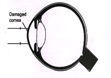
Corneal problems can happen to anyone at any age. Sometimes due to disease, injury or infection the cornea becomes cloudy or warped. A damaged cornea, like a frosted or misshapen windowpane, distorts light as it enters the eye. This not only causes distortion in vision, it may also cause pain.
When there is no other remedy, doctors advise a corneal transplant. In this procedure an ophthalmologist surgically replaces the diseased cornea with a healthy one to restore clear vision.


What is a Corneal transplant?
A transplant is the replacement of damaged or diseased tissues or organs with healthy tissues or organs. In a corneal transplant, the cloudy or warped cornea is replaced with a healthy cornea. If the new cornea heals without problems, there may be tremendous improvement in vision.
The healthy corneal tissue used for transplantation is supplied by an Eye Bank. Eye banks work round the clock to collect, evaluate, and store donated corneas. The corneas are collected from human donors within hours of death. Stringent tests are done to ensure the safety of the person receiving the cornea. The eye bank verifies the donor’s medical history and cause of death, and performs blood tests to ensure that the diseased person did not have any contagious disease, such as AIDS or hepatitis.
Since the cornea was one of the first parts of the body to be transplanted, corneal transplants remain one of the most common, and most successful, of all transplants.
How does the Eye work?
Anything you see is an image that enters your eye in the form of light. The different parts of your eye collect this light and send a message to your brain, enabling you to see. For perfect vision all the parts of your eye need to work properly.

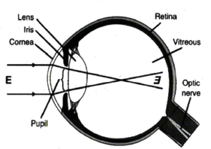
Light Rays passing through a healthy Cornea.
- The cornea is the clear, outer layer of the eye.
- The pupil is an opening that lets light enter the eye.
- The iris, the colored part of the eye, makes the pupil larger or smaller.
- The lens bends to focus light onto the retina.
- The retina receives light that has been focused by the cornea and lens.
- A clear (vitreous) gel fills the inside of the eye, giving it shape.
The Cornea is clear to let light into the eye, and curved to focus the light rays.
Some facts you may like to know:
- It is not necessary to find a cornea with a matching tissue or blood type.
- The race, gender and eye color of the donor are not important.
- A corneal transplant won’t change your natural eye color.
- The cornea heals slowly and improvement in vision may take a year or more.
- It is difficult to shape the new cornea perfectly. So, astigmatism (a condition where the cornea has an irregular shape, making images seem blurred or distorted) is common after a corneal transplant. However, this can be corrected.

Light Rays unable to pass through a damaged cornea.
Rejection of Transplant – the danger signals!
Rejection of a transplanted cornea can occur any time, but is more likely to happen in the first year after surgery. Unfortunately, rejection reduces the chances of success of any repeat corneal transplantation. However, this can be prevented by timely diagnosis and appropriate management.
Watch out for these danger signals:
- Redness
- Sensitivity to light
- Vision loss
- Pain
The acronym ‘RSVP’ can help you remember these symptoms. If you notice any of these symptoms in your operated eye, however minor they may seem and regardless of the time of day, contact us immediately. If this is not possible, visit nearest ophthalmologist, preferably a cornea specialist.
As with other surgical procedures, a corneal transplant involves some risks – most of them can be treated. Some possible complications are :-
- Eye infection
- Failure of the donor cornea to function normally
- Rejection of the donor cornea by your body
- Cataract (clouding of the eye’s lens)
- Glaucoma (build-up of fluid, leading to increased pressure in the eye)
- Bleeding from the iris
- Swelling or detachment of retina
Important Tips on Care after Surgery
You can bathe carefully from below your neck, and also shave, but do not let the operated eye become wet for at least 15 days. You may gently clean the eyelids with a piece of cotton boiled in water or a sterilized tissue. Do not wet the eyeball. You should wear an eye patch at night; the doctor will advise you when to discontinue using it during the day. Always wear protective glasses or an ‘eye shield’ to avoid accidental injury.
- Do not lift heavy things
- Do not bend so that your head is lower than your waist
- Avoid sleeping on the operated side
- No sexual intercourse until permitted by the doctor
- Do not rub the operated eye
- Avoid any vigorous activity
- Avoid alcoholic beverages
- Watch television for short periods only
Medication and Follow-up
At the time of discharge our patient counselor will advise you about medication and follow-up visits. Please follow the instructions regarding medication. Please adhere to the follow-up appointment date.
If you have any concerns or questions, you can ask the doctor when you can come for an examination. If you feel you cannot wait, call or email us, or send a fax, at our numbers given.
If there is an emergency at night, during a weekend, or on a holiday, come for emergency care to the institute.
Always mention the patient’s ID number, name and the doctor’s name in all communications.
