Anatomy of the Eye
- Education
- Anatomy of the Eye
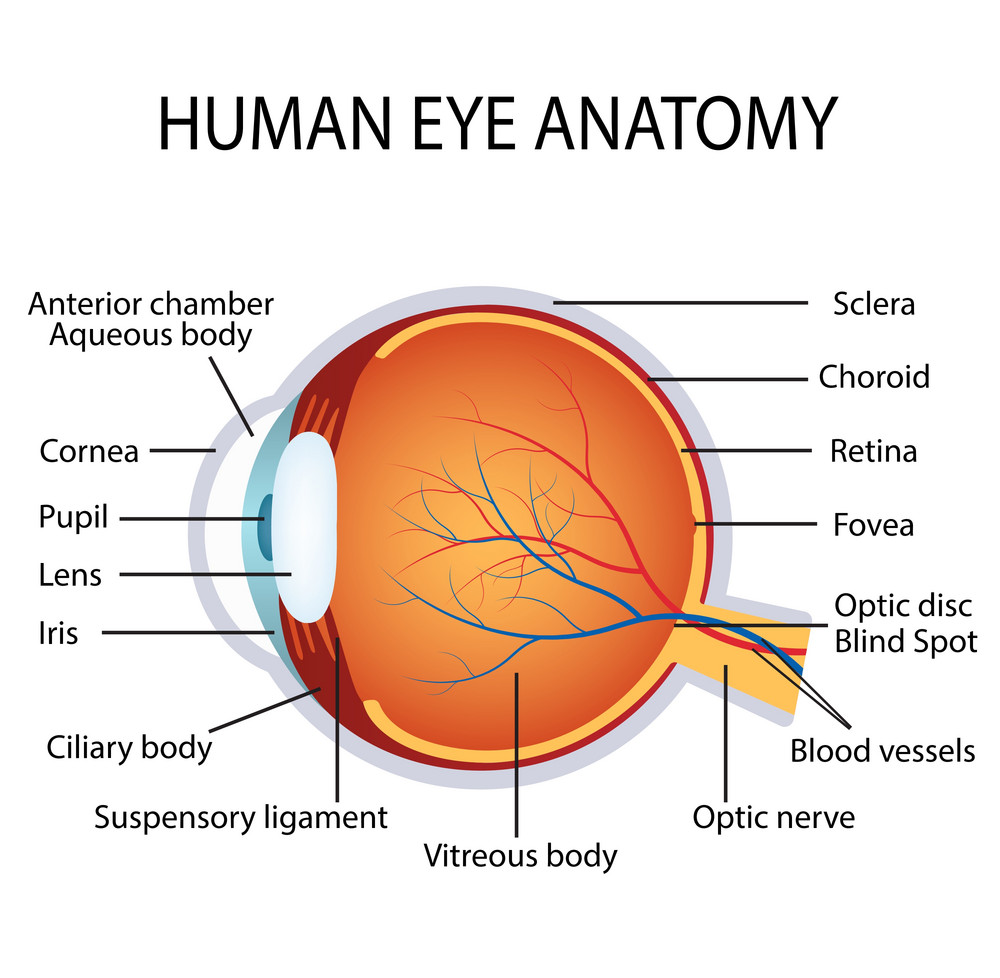
A clear gel that fills the eye.
A bundle of more than one million nerve fibers that carry visual messages from the retina to the brain.
The small, sensitive area of the retina that gives central vision. It is located in the center of the retina and contains the fovea.
The center of the macula; it gives the clearest vision.
The light-sensitive tissue lining at the back of the eye. The retina converts light into electrical impulses that are sent to the brain through the optic nerve.
The colored part of the eye that regulates the amount of light entering the eye.
A clear part of the eye behind the iris that helps focus light or an image on the retina.
The opening at the center of the iris. The iris adjusts the size of the pupil and controls the amount of light that can enter the eye.
The cornea is the transparent circular part of the front of the human eyeball.
It has an important optical function as it refracts light entering the eye through the pupil and onto the lens which then focuses the light onto the retina.
Cornea a non-vascular structure which means that it does not contain any blood vessels as the capillaries that supply it with nutrients terminate in loops at its circumference. It is supplied by many nerves derived from the cilliary nerves. These enter the laminated tissue of the cornea. It is therefore extremely sensitive.
It borders with the sclera by the corneal limbus.
The human cornea has five layers; From the anterior to posterior the five layers of the human cornea are:
- Corneal Epithelium.
- Bowman’s Layer.
- Corneal Stroma.
- Descemet’s Membrane
- Corneal Endothelium.